


Which is a Magnetic Resonance? is a non-invasive and painless procedure that provides a very detailed view of the structure and composition of the tissues to be analyzed. Magnetic Resonance does not use X-rays, but magnetic fields to build images that help detect congenital, infectious, traumatic, vascular, tumor or degenerative alterations in any part of the body.
It is essential in the analysis of neurological conditions such as epilepsy, heart attack and dementia. Also in the study of osteoarticular pathologies and in the evaluation of the thorax, abdomen, pelvis and vascular system.
MRI provides a clearer view of the interior of the body compared to other techniques and reduces the need for certain diagnostic surgeries. It may be necessary to use intravenous contrast dye to better visualize structures or fluids within the body.
We provide the MRI service in Medellín at Calle 59 No. 50 A 14 (Prado Centro neighborhood) and in the municipality of Sabaneta at Carrera 48 No. 50 Sur 128 (Mayorca Mega Plaza Shopping Center).
Take advantage of our discounts and benefits on private exams!
Request your private appointment on WhatsApp 316 403 76 61 (chat only).
Special rates.
Opportunity in assigning appointments.
Results in two business days.
The patient must not have metallic elements in his body due to the high intensity magnetic fields. If it is necessary to apply the contrast medium, the nurse will channel a vein.
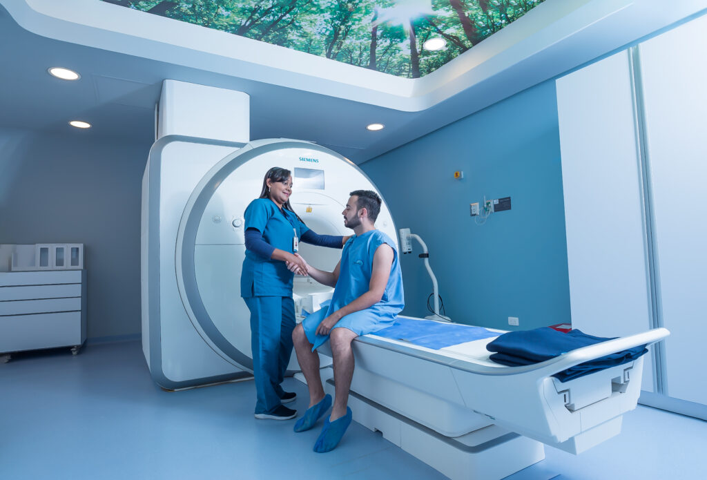
The Diagnostic Aid Technologist or nurse will help you lie down on the equipment table, usually on your back. The area of the body that will be subjected to MRI must be in the center of the resonator where the magnet is located, which will emit loud sounds while the images are being taken.
The technologist will remain in contact with the patient throughout the duration of the exam through an audiovisual system, and will indicate the exact moments in which they must remain immobile to guarantee the quality of the images and prevent them from coming out blurry. In addition, for certain diagnoses, it will ask you to hold your breath for brief moments.
MRI does not cause any pain or side effects. However, when contrast media is administered, in some cases the patient may experience nausea, flushing, a sensation of heat, and/or headache. Sometimes a patient may experience an allergic reaction, for which we have a permanent doctor who evaluates and treats this type of adversity. If the patient ever feels anything strange, it is important to report it to the technologist or nurse immediately.
The duration of the exam varies according to the part of the body to be examined, although the approximate average time is thirty (30) minutes.
The images are recorded on a CD, preserving the entire study. Additionally, the interpretation of the Radiologist is delivered.
- Magnetic resonance imaging of the pelvis.
- Magnetic resonance imaging of the cervical, lumbar or thoracic spine.
- Magnetic resonance imaging of joints and extremities.
- Nuclear magnetic resonance of the brain and advanced techniques: spectroscopy, diffusion, perfusion, tractography and functional BOLD.
- Magnetic resonance imaging of the temporomandibular joint (TMJ).
- Abdominal magnetic resonance imaging.
- Chest magnetic resonance imaging.
- Neck magnetic resonance imaging.
- Sella turcica magnetic resonance imaging.
- Orbital magnetic resonance imaging.
- Magnetic resonance imaging of the ear.
- Magnetic resonance imaging of the shoulder.
- Angioresonance.
- Arthroresonance.
- Enteroresonance.
- Simple resonances: pelvis - cervical, lumbar or thoracic spine - brain - TMJ - abdomen - thorax - orbits - ear - joints and extremities.
- Contrasted resonances: pelvis - cervical, lumbar or thoracic spine - brain - joints and extremities - shoulder - arthroresonance - angioresonance - TMJ - abdomen - thorax - orbits - ear - neck - sella turcica.
- Contrasted entero-magnetic resonance.
- MRIs under sedation for adults and children over two years of age.
- MRIs under sedation for children under two years of age.
- Contrast-enhanced multiparametric prostate resonance.
- contraindications.
- Contrast MRIs: Abdomen, Uroresonance, Cholangioresonance.
3.0 Tesla Magnetom Verio Resonator – Siemens
- Equipment with the highest power and the best quality for morphological and functional studies with excellent resolution.
- Tim technology (Total imaging matrix) that expands the potential of the 3T.
- It allows highly specialized studies such as tractography of the central nervous system, brain functional studies, magnetic susceptibility imaging, cerebrospinal fluid dynamics, conventional and time-resolved angioresonance studies, spectroscopy (uni-multivoxel, 2D-3D), diffusion and prefusion ( with and without contrast), having the highest spatial resolution possible.
- Comfort and biosecurity for the user.
- Short and wide tunnel for patients with obesity or claustrophobia.
- It has applications for orthopedics, body image, neurological images and vascular studies.
- Latest technological innovations that allow the interconnection of the same to a computer network of images.
1.5 Tesla Magnetom Aera Resonator – Siemens
- Superior image quality for excellent diagnostic accuracy.
- Shorter studies depending on the type of exam.
- High quality and exceptional speed with TIM 4G.
- It has a wide range of clinical applications that allow the 1.5 Tesla technology to be maximized.
- Better access for patients with motor difficulties, the elderly and children.
- Reduces sound pressure by up to 99% to improve the patient experience.
1.5 Tesla Magnetom Essenza Resonator – Siemens
- 1.5 Tesla Field: Provides excellent image quality.
- Complete range of clinical applications: Neuro, Ortho, Body, Angio, Onco and Pediatric.
- Compact size.
- Ultra short exploration tunnel, 145 cm. It allows studies with the head and feet of the patient outside of it, avoiding the feeling of claustrophobia.
- Diagnostic time lower than other similar equipment.
- Coils that make it possible to carry out studies in all parts of the human body in adults and children.
- Its light coils allow more comfortable studies for the patient.
- The equipment has the latest technological innovations that allow its interconnection to an image computer network.